Raman spectrometer – Renishaw inVia Qontor confocal microscope 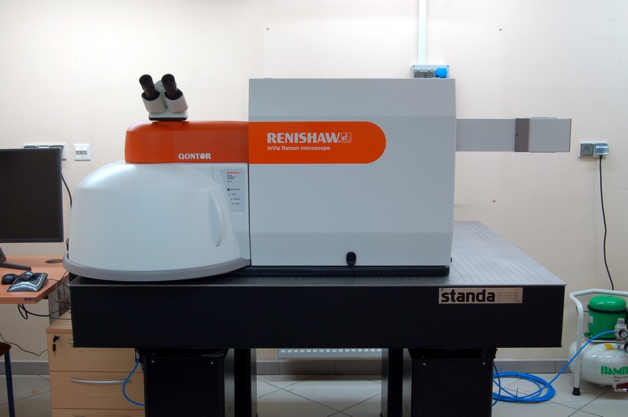 The inVia Qontor system is a spectrometer used for studying the physicochemical and molecular properties at the nanoscale. The principle of operation of the device involves analyzing the scattering of a laser light beam by chemical molecules present in the sample being studied.
Most of the light scattered by the molecules has the same wavelength as the incident light (known as Rayleigh scattering), but a very small amount (about ten million times less than Rayleigh scattering) has a changed wavelength due to the influence of molecular vibrations in the sample on the scattered light (known as Raman scattering). It is this additional, modified light that provides information about the vibrations of chemical bonds in the substances present in the sample.
The recorded information on the intensities of the scattered light for different wavelengths is called a Raman spectrum. Based on such spectra, and using appropriate libraries, it is possible to identify substances, their crystalline forms, estimate the composition of mixtures, or detect mechanical stresses present in the substance. It is also possible to analyze the sample point by point with a step size reaching one hundred nanometers (one ten-millionth of a meter). Pure metals cannot be studied using the Raman spectroscopy method, and some bond vibrations in non-metallic substances may require complementary infrared spectroscopy.
The inVia Qontor system also enables surface imaging of the sample using Rayleigh scattering (known as Rayleigh mapping). These measurements are very quick because Rayleigh scattering provides much stronger radiation than Raman scattering, making it technically easier to perform.
The measurement setup consists of a confocal Raman microscope and a PC that controls its operation using the dedicated WIRE application, which runs in the Microsoft Windows environment. This application provides measurement results in *.wdf file format, and some specific data can be saved in standard bitmap image formats or as text files.
The equipment is registered in the Laboratory of Materials Research Engineering at the University of Zielona Góra, in the part belonging to the Institute of Physics.
Basic features of the inVia Qontor confocal Raman microscope:
Available lasers: 532 nm, 785 nm
Magnifications of available microscope objectives: 5x, 10x, 50x, 100x
Range of motion of the motorized microscope stage: 112 mm (x-axis), 76 mm (y-axis), 25 mm (z-axis)
Centrus detector (CCD technology, range 200 nm – 1064 nm, operating temperature -70°C)
Nanosurf Flex-Axiom Atomic Force Microscope
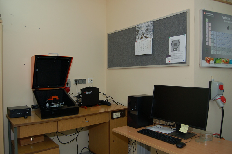 An atomic force microscope (AFM) is a device that allows the study of physical phenomena occurring at the nanometer or micrometer scale on material surfaces. The principle of operation is based on analyzing the forces between the material's surface and a probe tip that is scanned over a designated area of the surface. The simplest application of an atomic force microscope is surface imaging. This is achieved by monitoring the height adjustment (z-axis) of the probe suspension above the studied surface, necessary to maintain a predetermined interaction parameter between the probe and the surface of the analyzed material (e.g., the force of the probe pressing on the surface). In this way, information about the height of the sample at a given point on the material is obtained. However, other specialized measurements are also possible, such as measuring magnetic forces or surface conductivity, using specialized probes.
The measurement setup consists of the atomic force microscope and a PC that controls its operation using the dedicated Nanosurf C3000 application, which runs in the Microsoft Windows environment. This application provides measurement results in *.nid file format, and some specific data can be saved in standard bitmap image formats.
Properties of the Flex-Axiom atomic force microscope:
Maximum scanned area size (x and y axes): 100 x 100 micrometers
Maximum height range (z-axis): 10 micrometers
Keyence VHX-970F Digital Microscope 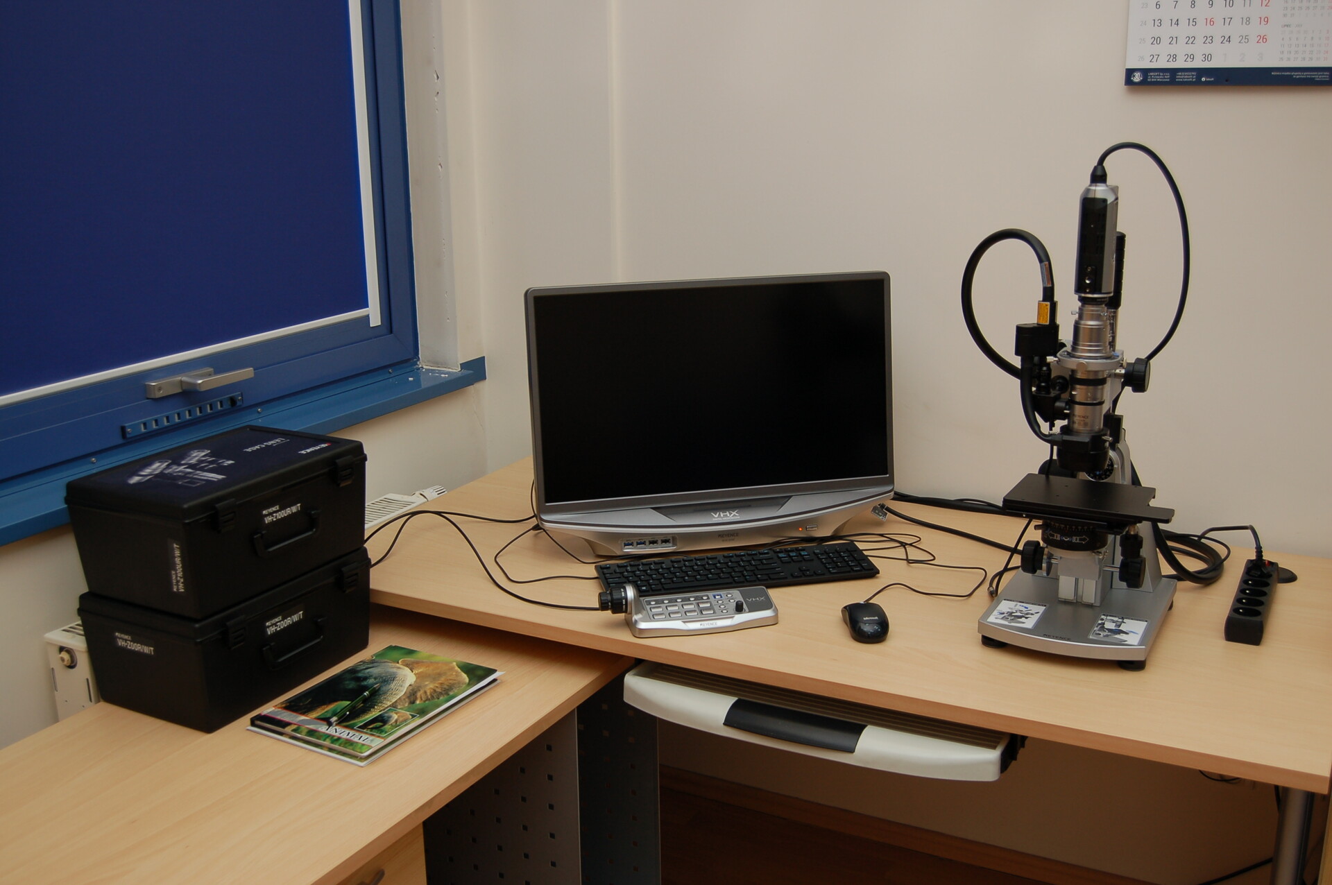 A digital microscope is used for observing miniature objects at magnifications ranging from several times to a thousand times, with the capability of digitally recording the obtained images at a resolution of 2048x1536 pixels (3 MP). Static images and videos are saved in typical, popular file formats (JPEG, TIFF, AVI). The optical system of the microscope allows for the adjustment of lighting in a wide range in terms of the direction and intensity of the incident light, depending on the needs. The microscope software enables the measurement of the geometry of observed objects. Advanced functions are also available, such as eliminating reflections on the surfaces of observed objects. The combination of digital technology with automatic focus control of the lens has additionally allowed the manufacturers to provide users with functions such as imaging with a wide depth of field and creating three-dimensional images of observed objects.
Equipment included:
Wide-angle zoom lens with a magnification range of 100x-1000x
Variable-focus lens with a low magnification range of 5x-50x
VHX-A97FP control console
Shimadzu UV-2600 Spectrophotometer 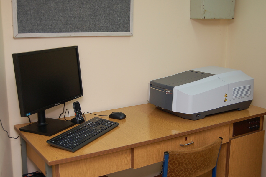 A spectrophotometer is a device used to study the optical absorption of substances. It enables the determination of how the amount of radiation transmitted through a sample changes as a function of its wavelength. Such information allows for determining the optical properties of transparent materials (transmission range, attenuation), identifying optically active substances in solids and solutions, including the oxidation states of dopant ions. Another common application of the spectrophotometer is determining the concentrations of substances in solutions by comparing the optical absorption of the test solution with the optical absorption of a series of standard solutions with known concentrations of the target substance.
The measurement setup consists of the spectrophotometer and a PC that controls its operation using the dedicated UVProbe application, which runs in the Microsoft Windows environment. This application provides measurement results in its internal formats, dedicated to various types of measurements, and allows data export to a text file.
Properties of the UV-2600 spectrophotometer:
50W halogen lamp and deuterium lamp, automatically switched
Wavelength range from 185 nm to 900 nm
Absorbance measurement range from -5 to 5
EPR spectrometers
The Electron Paramagnetic Resonance (EPR) spectrometer is used to study paramagnetic substances by recording the absorption of microwave radiation as a function of an applied external magnetic field. Paramagnetic substances contain paramagnetic centers, which are atoms, molecules, or molecular complexes with uncompensated spin. These paramagnetic centers can include free radicals, transition metal ions, rare earth ions, or defects in dielectric materials. The EPR technique can provide valuable information about the paramagnetic centers in a substance, such as the oxidation state of dopant ions or the geometry of their local environment.
X-Band EPR Spectrometer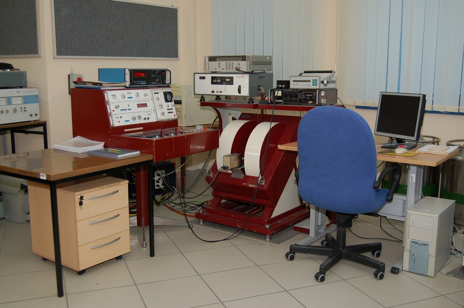
The EPR measurement setup in the X-band microwave range consists of the Radiopan SE/X-2013 spectrometer along with additionalequipment: an HP 5350 frequency counter from Hewlett-Packard, a 20 NMR magnetometer, a dedicated signal digitization module, and a PC used for spectrum recording. The application for spectrum recording operates in a Linux environment and provides data in text file format.
Technical Specifications of the Radiopan SE/X-2013 Spectrometer:
Klystron: 100 mW power, typical operating frequency around 9.4 GHz, frequency stability 10^-6, microwave cavity quality factor Q0=7000
Electromagnet: Operating range 0-880 mT, field sweep time range 1-100 minutes, sweep non-linearity 1%
Magnetic Field Modulation Frequency: 100 kHz
 The EPR measurement setup in the Q-band microwave range consists of the EPRAD-RADIOFAN SE/X-2547 spectrometer. Technical Specifications of the EPRAD-RADIOFAN SE/X-2547 Spectrometer:
Gunn Diode: 100 mW power, operating frequency 34 GHz
Electromagnet: Operating range 0-1440 mT, field sweep range 1000 mT
Magnetic Field Modulation Frequency: 100 kHz
FluoroMax-4 Spectrofluorometer by Horiba Jobin Yvon 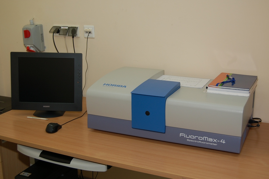 A spectrofluorometer is a device used to study the phenomenon of luminescence in substances. It works by emitting radiation from a substance when it is illuminated, with the emitted radiation having longer wavelengths than the excitation radiation that stimulates the substance. Characterizing the luminescence of a substance involves recording excitation spectra, emission spectra, and determining the kinetics of luminescence decay. The FluoroMax-4 spectrofluorometer allows for the performance of all these tasks.
The measurement setup consists of the spectrofluorometer and a PC that controls its operation using the dedicated FluorEssence application, which runs in the Microsoft Windows environment. This application provides measurement results in *.opj format, allowing for subsequent analysis using Origin software by OriginLab.
Properties of the FluoroMax-4 Spectrofluorometer:
Xenon Lamp: 150 W, with a wavelength range of 240-850 nm
Excitation Monochromator: Adjustable for the wavelength range of 220-600 nm
Emission Monochromator: Adjustable for the wavelength range of 290-850 nm
Photomultiplier Tube: R928P by Hamamatsu, operating in single-photon counting mode, sensitive in the wavelength range of 200-850 nm
Luminescence Lifetime Measurement Range: Minimum of 10 microseconds
RADWAG WPA180/K Analytical Balance 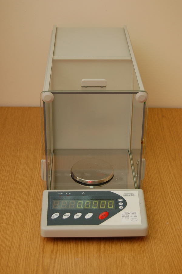
Technical Specifications:
Capacity: 10 mg - 180 g
Readability: 0.1 mg
Tare Range: -180 g
Protherm PTF 12/38/250 Tube Furnace 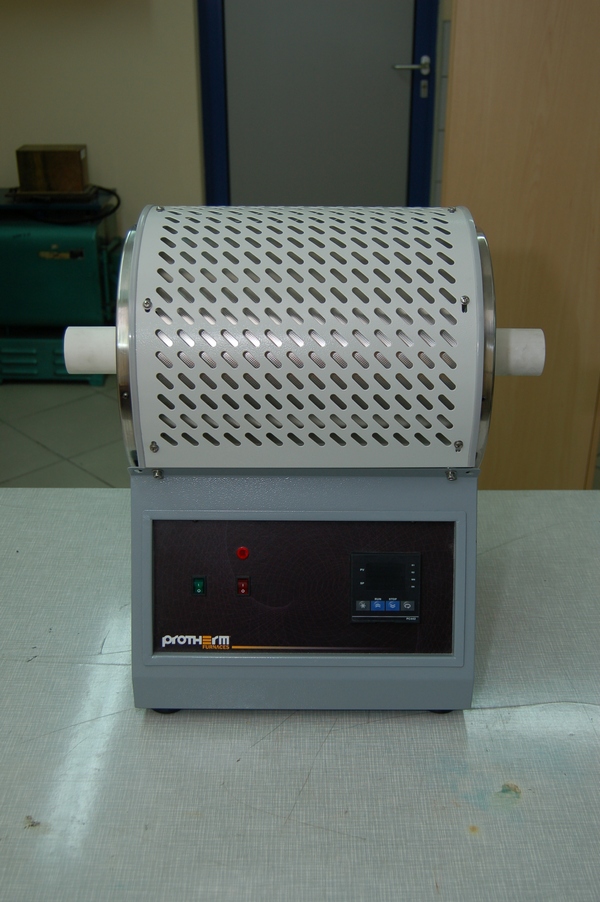
Technical Specifications:
Tube Size: 38x500 mm
Heated Zone: 250 mm
Maximum Temperature: 1200 °C
Temperature Deviation: ±2 °C
Power: 1.3 kW
MPW Med. Instruments MPW-251 Centrifuge 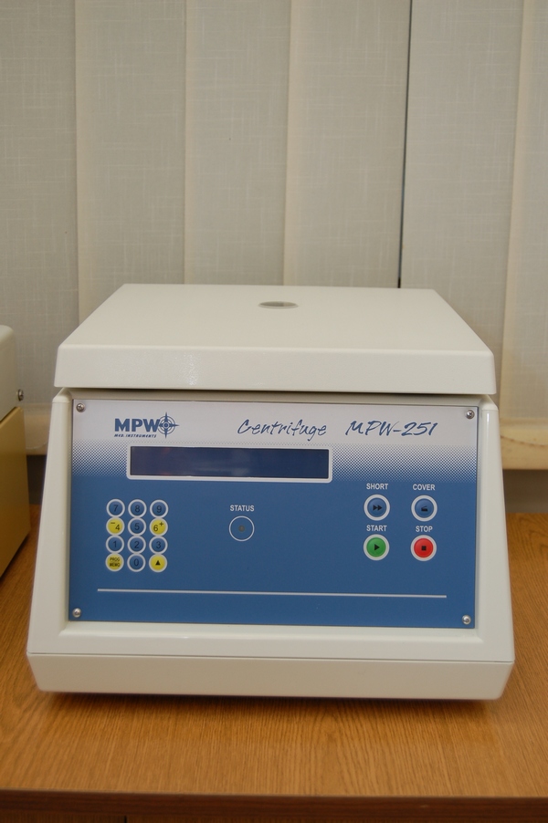
Technical Specifications:
Rotation Speed: 100-18,000 rpm, increment 100 rpm
Relative Centrifugal Force (RCF): 24,270 g
Centrifugation Time: 0-99 minutes 99 seconds, increment 1 second, or continuous operation
Additional Equipment Includes:
Angle Rotor 12x1.5/2 ml: 18,000 rpm, RCF: 24,088 g, 45 degrees
Angle Rotor 6x50 ml: 6,000 rpm, RCF: 4,427 g, 30 degrees
Fume Hood 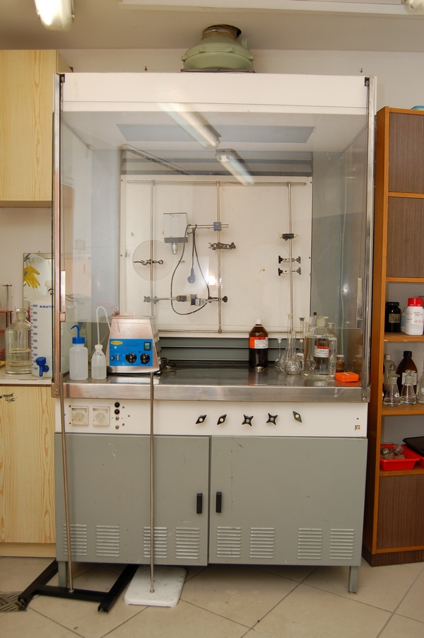
Leica ACE200 Low-Pressure Coater 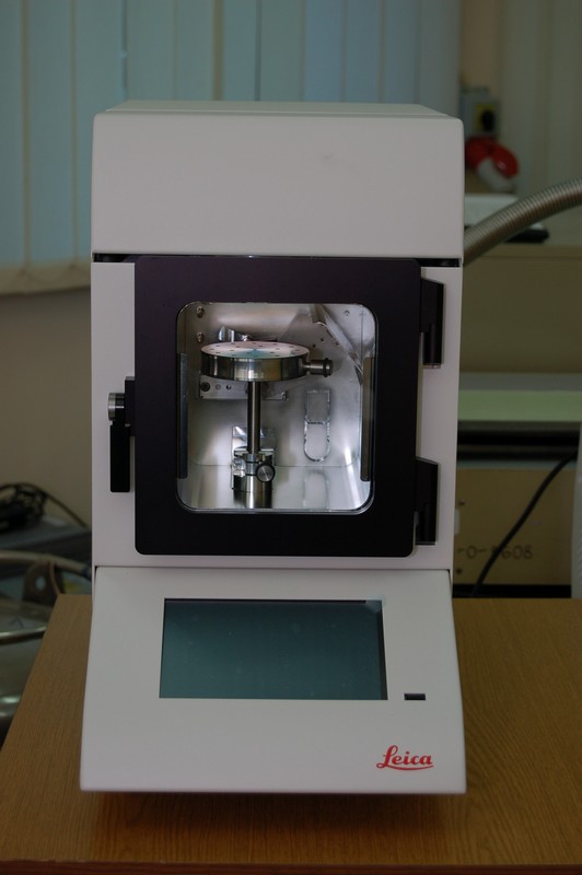
Leica ACE200 Low-Pressure Coater for Producing Thin Carbon Layers
FDM 3D Printer (Filament-Based), ZIG-ZAG 3D Series 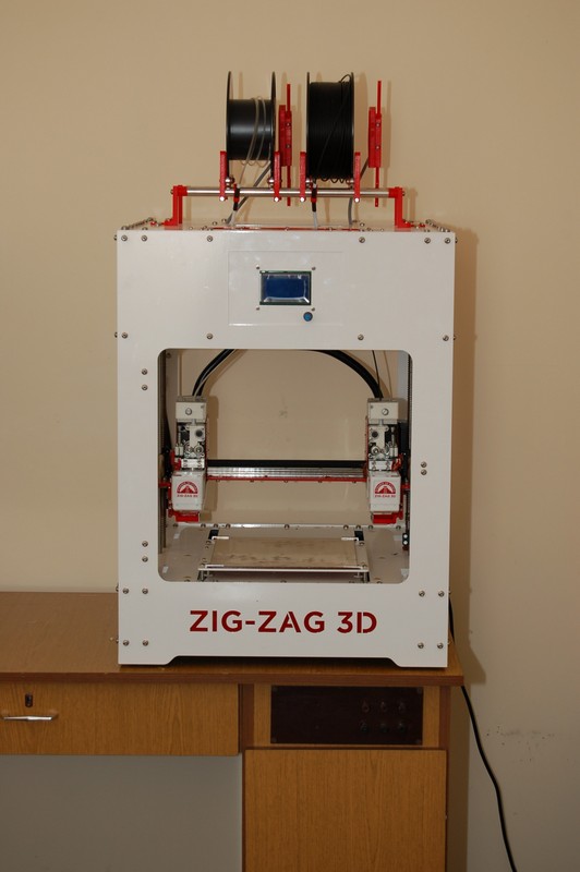
FDM 3D Printer (Filament-Based), ZIG-ZAG 3D Series – Available at the Laboratory of Material Research Engineering, University of Zielona Góra, in the section belonging to the Institute of Physics.
SLA 3D Printer, Formlabs Form 3 Series 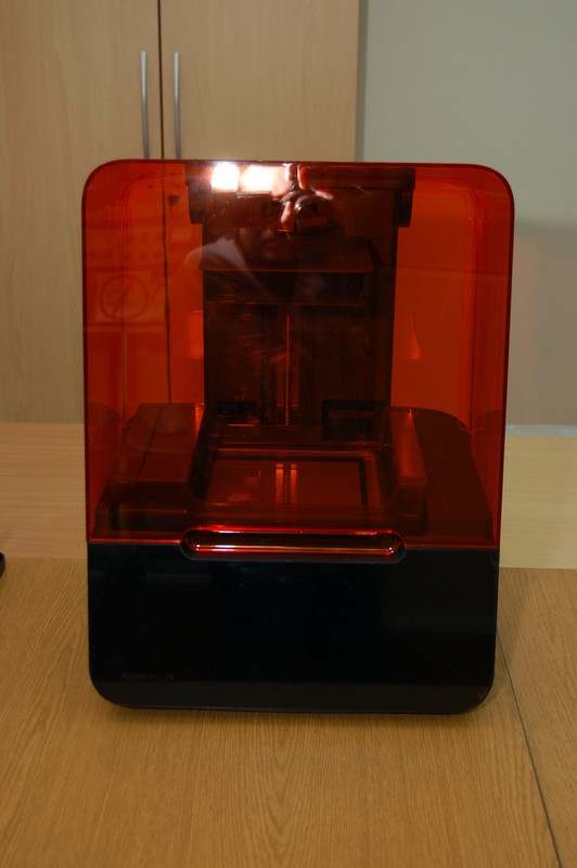
SLA 3D Printer, Formlabs Form 3 Series – Available at the Laboratory of Material Research Engineering, University of Zielona Góra, in the section belonging to the Institute of Physics.
Malvern Dynamic Light Scattering Particle Size, Colloid Stability, and Zeta Potential Analyzer
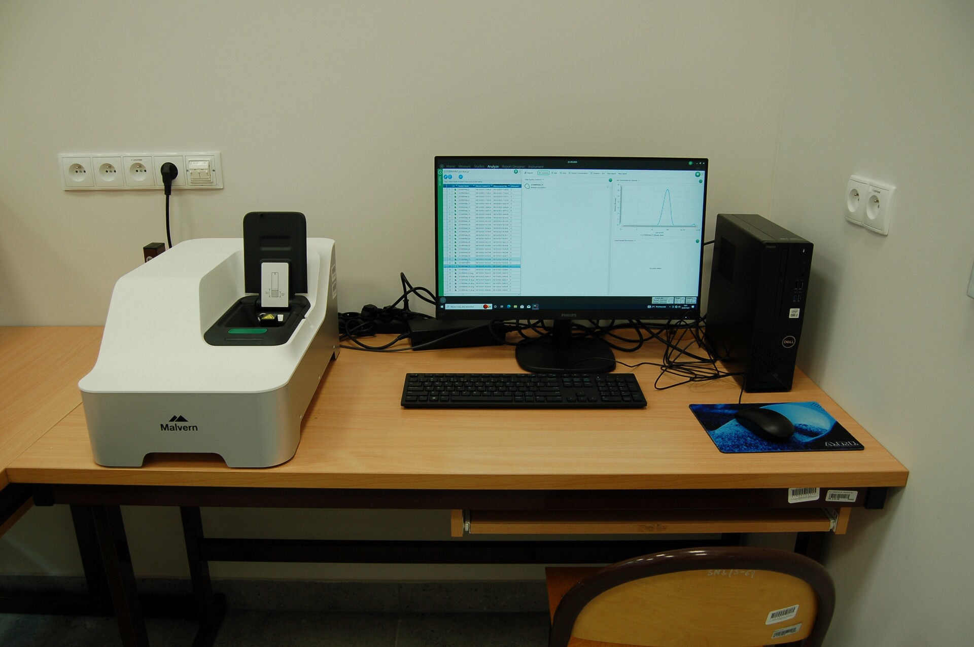 The equipment is available in the Materials Research Engineering Laboratory at the University of Zielona Góra, within the Department of Physics. This apparatus is used for assessing the properties and quality of nanoparticles synthesized in the Chemical Laboratory of the Department of Physics.
Glovebox
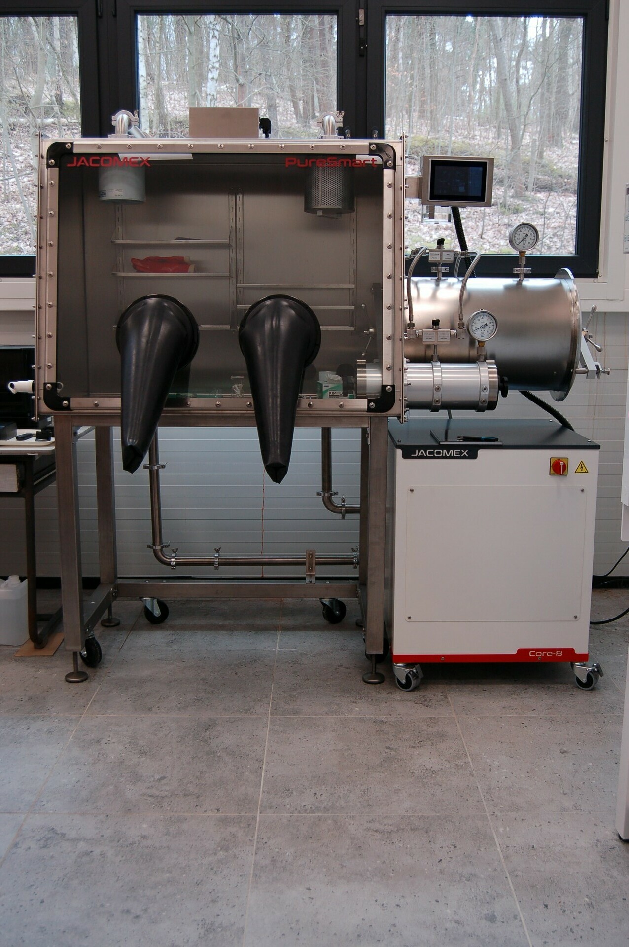 The equipment is available at the Laboratory of Material Research Engineering, University of Zielona Góra, in the section belonging to the Institute of Physics